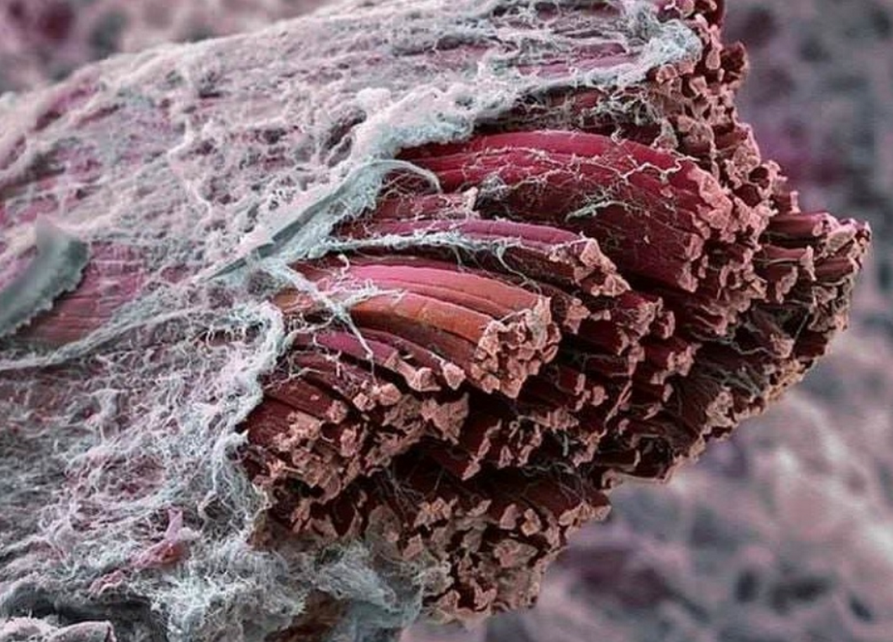
Fascia, fascia, fascia – many talk about it, they supposedly seem to be good for many things and responsible for various zippers. The fitness industry rolls over with “fascial” offers. But what is fascia anyway? Where are they in the body? What characteristics and tasks do they have?
The non-vegetarians/non-vegans among us who pursue the passion of cooking have certainly already seen, touched and cursed fascias. For example, they envelop muscle meat – very nice to see with a piece of chicken breast – as a milky white or silvery shimmering thin membrane.
When trying to remove this shell, meat fibers are more likely to be pulled out, because the shell seems to continue inside the meat. And in some places it thickens into a solid strand, the removal of which requires some patience even with a sharp knife.
All those who do not eat meat may forgive me for the comparisons and can look forward to a different picture below. But what has been described for the muscle meat of animals also applies to the human muscles: they are enveloped and permeated by fascia.
A question of definition
Let’s start a little more formally. The word fascia is derived from the Latin word “fascia”, which means bandage, ribbon or bundle. In German, one would speak of connective tissue. This seems kind of familiar to you, but rather leads to misunderstandings.
Medically speaking, bones and blood belong to the connective tissue, among other things, because when dividing the tissues, only the cell properties, the cell spaces and the embryonic development history are looked at here. Bones and blood, however, are not the subject of fascia research, although the former are essential for understanding the movement function and, in addition, the bone skin (the perioost) cannot be pulled out of the fascia world so clearly. The outer bone skin layer (stratum fibrosum) is connective tissue-like.
So anatomy recently agreed on the following great definition:
A fascia is a sheath, a sheet, or any other
Dissectable aggregation of connective tissue that
Forms beneath the skin to attach, enclose, and
Separate muscles and other internal organs’
Sounds complicated. If you look closely, you will discover the envelopes and the word “dissectible”. Anatoms like to prepare something clearly to name and describe it. This definition would not include tendons, ligaments and joint capsules and would not be conducive to the holistic view of a health and movement orientation even against the background of the division into small sections.
That’s why the fascia researchers have also provided a so-called functional conceptualization, which defines the fascia system as a coherent network of fibrous, collagen connective tissues, the shape and structure of which is decisively determined by tensile forces.
No less complicated. For our considerations, the words network, collagen fiber and tensile forces are decisive, as will be shown below. Muscular connective tissue, tendons, ligaments, joint capsules are among the fascias, which suggests the importance for the mobility of our body.
Where can we find fascia in the body?
Almost everywhere.
But we don’t want to make it that easy for ourselves. Too complicated, however, neither would it be, which is why the following personal subdivision has more practical reasons and would not withstand a scientific discourse.
- The skin covers our body and under the epidermis lies the first loose fascia superficialis layer that houses the subcutaneous fat tissue – the outermost shell, so to speak. It ensures firmness of the skin and mobility to the underlying structures. Whether one can actually speak of a uniform fascial class is scientifically controversial.
- Below lies the deep fascia layer (fascia profunda) as a firmer second shell. It gives shape to the body – a cover, like a diving suit.
- Below are the muscles, individual muscle bundles, even the muscle fibers packed in fascia. The muscular connective tissue (myofascia) prolongs and thickens in tendons, form flat structures, come to light here and there thicker, firmer, are interwoven with each other and with ligaments and capsules, but equally separated from each other.
- Ultimately, our organs, organ groups are also enveloped and padded (visceral fascia) and even nerves and brains have their packaging material (meningeal fascia).
The structure should not deceive over the following facts: Fascias are woven and connected everywhere, merge into each other and the individual envelopes refine into ever smaller units – almost like the skins inside a lime.


This makes it clear why the above definition speaks of a network. And it makes it clear that problems at one point on the network can potentially lead to complaints in a completely different place.
Tensegrity
With this network idea, we come back to the bones. These are firm and pressure-stable, while the fascias are more oriented towards tensile stress. A structure that embeds pressure-stable components in a coherent voltage network without touching each other is called a Tensegrity structure. The word derives from “tension” and “integrity”. The invention or rather discovery or description is attributed to Richard Buckminster Fuller, an architect and designer.
If you transfer the model, the human body would be a complex Tensegrity structure, which again shows how tension changes (increased muscle tone, scars, ligament stretching, etc.) can have an effect in completely different places of such a structure. And it would have the interesting consequence that the pressure-stable components, i.e. the bones, do not touch at optimal tension. An interesting idea that we are not a stack of bones, but “hang” the bones in the fascial system. The spine would not be a column, but a system of tense individual vertebrae. The concept of joint gap shows that this is not absurd.
But caution is advised. It’s a model. The proof that the body is actually a tensegrale structure macroscopically has not (yet) been scientifically successful. At the cellular level, on the other hand, Tensegrity laws seem to apply. Back from our excursion…
What are fascias made of?
From water. About two-thirds. Even if we dry out a little in the course of our lives, it is amazing that water is supposed to keep us in shape. The water is bound by sugar-protein compounds: proteoglycans and glycosaminoglycans for the chemists among us. If you can’t imagine anything about it, you may have heard of hyaluronic acid before, more right: Hyaluronan. Only one gram of it can bind up to 6 liters of water. Water and hyaluronic acid act (if in the right proportion) like lubricants and ensure the mobility of the fascial layers.
In this slippery mixture we find elastic fibers: essentially made of collagen (protein). These fibers are arranged differently depending on the function of the fascial fabric and influence the mechanical properties.
All together is called extracellular matrix, which indicates that cells are embedded in the fascia. Although these account for only 8-9% of the fascial tissue, they have it all. The so-called fibroblasts (in inactive form fibrocytes) are the builders of the fascia. They generate and destroy the components of the extracellular matrix. Immune cells provide for our health, foothills of nerve cells for perception.
And what does this have to do with exercise?
It is fascinating that despite the same “ingredients”, the different composition and structuring of the fibers leads to very different properties.
In the envelopes around the muscles, we often find a scissors grid-like arrangement, which makes it possible to give in to its thickening when tightening a muscle group. The covers have a certain elasticity and a “watery” layering, which makes movements possible in the first place.
The elasticity is not so great along the muscle fibers – here the focus is on the transmission of power. Especially in the transition to and in the tendons, where the fibers are arranged rather parallel and form a kind of firm pull rope.
In healthy fascias, the fibers also have a wavy structure. This, in combination with the interdependence and interaction with the other components of the extracellular matrix, leads to visco-elastic behavior: a fascial structure can transmit tensile forces and it can absorb kinetic energy and release it quickly, which makes our walking, running, jumping, throwing, etc. so efficient. But it reacts, as it were, with deformation, which is only slowly lifted again, similar to a long gummy bear. We look at these properties in detail in subsequent articles.
But that’s not all
The connection to the nervous system has already been mentioned above. Countless (mechano) receptors are embedded in the fascial tissue, which make it a rich source of information. The fascia system is our largest sensory organ and one could speak of the sixth sense. In technical terms: proprioception. (Basically also interoception in the narrow sense, but also more about it in a separate article.)
Proprioception is the self-perception of body movement and position in space and is mediated by the equilibrium organ in the inner ear and by the different mechanoreceptors. Without proprioception, no targeted, safe, fine, elegant, emotional movement would be possible.
So our fascias have a great influence on our mobility. But vice versa, they also react to movement: the arrangement of the fibers in the extracellular matrix is carried out by movement and the fibroblasts also react to mechanical stimuli with the removal or build-up of extracellular matrix. And we will continue this line of thought in the next article on the trainability of the fascial system.


